Handling the Lenticonus Challenge

Alport syndrome is a multisystem disorder characterized by a defect in collagen IV synthesis, resulting in progressive nephritis and sensorineural hearing loss with multiple ocular abnormalities of which the anterior lenticonus is pathognomonic. (1).

1
The Background

This case report emphasises the importance of accurate diagnosis of anterior lenticonus and the crucial role of an ophthalmologist in aiding the timely diagnosis of Alport syndrome.

  • A 16 year old boy, with progressively decreasing vision and BCVA 6/36 OD and 6/24 OS, came in for opinion about CXL.
  • Distant direct ophthalmoscopy showed a central symmetrical oil-droplet sign
  • Slit lamp exam showed conical protrusion of anterior lens capsule and a normal posterior segment.

Figure 2: Oil droplet reflection in retro-illumination (indicated by arrow on left. Cross-section showing protrusion of anterior lens capsule (highlighted by bold arrow on right).

  • The progressive bulging of the capsule creates a central, symmetrical oil droplet appearance on retro-illumination which can mimic keratoconus. However, slit lamp examination and A/S OCT can omit this confusion.
  • The structural fragility of the capsule gives rise to a cogwheel tearing pattern, which accounts for a challenging capsulorrhexis with risk of rips and runoffs (2). Meticulous care should be taken during capsulorrhexis in these cases.​
2
The Workup
  • Anterior Segment OCT
  • Urinalysis
  • Pure Tone Audiometry

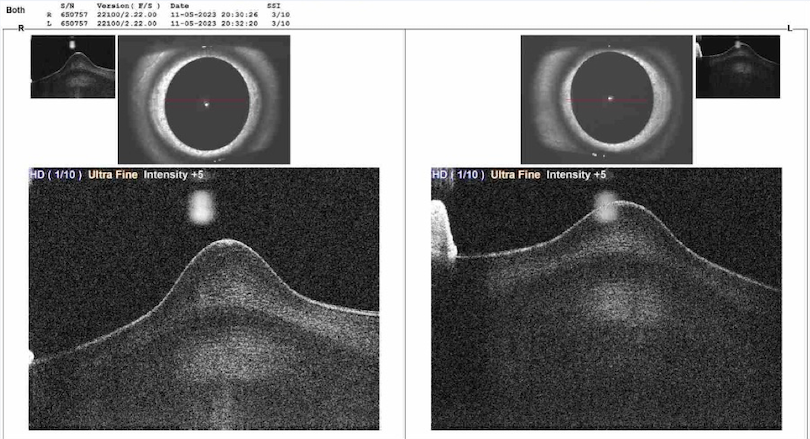
Figure 1: Anterior Segment OCT demonstrating Bilateral Anterior Lenticonus.

3
The Challenges
4
The Plan Exercised
  • Irrigation and aspiration with IOL implantation OU.
  • Referral for auditory rehabilitation and renal evaluation.

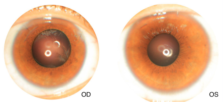
Figure 3: 4 weeks post-op slit lamp photograph with BCVA of 6/9 OU.

5
The Triumphs

Anterior lenticonus of Alport Syndrome can be successfully managed with clear lens exchange and IOL implantation. Early recognition facilitates timely diagnosis, and administering ACE inhibitors and ARBs can delay end-stage renal disease.

Frequently Asked Questions (FAQs)

What are the primary ocular signs of Alport syndrome, and how are they diagnosed?
Alport syndrome commonly causes anterior lenticonus. We diagnose it using slit-lamp examinations and Anterior Segment OCT to identify the characteristic conical lens protrusion and oil-droplet appearance.
How is anterior lenticonus differentiated from keratoconus?
Anterior lenticonus affects the lens, while keratoconus affects the cornea. Slit-lamp exams and Anterior Segment OCT at Acuity Eye Center distinguish the lens's conical protrusion in anterior lenticonus from corneal changes in keratoconus.
What treatments are available for anterior lenticonus at Acuity Eye Center?
We treat anterior lenticonus with clear lens exchange and intraocular lens (IOL) implantation. We also refer patients for auditory and renal evaluations due to the multisystem nature of Alport syndrome.
What surgical precautions are taken for Alport syndrome patients?
Our surgeons take extra care during capsulorrhexis to avoid capsule tearing. The fragile lens capsule requires precise techniques to prevent complications during lens exchange surgeries.
How does early treatment affect Alport syndrome prognosis?
Early treatment improves vision and delays renal disease progression. Timely interventions at Acuity Eye Center, including lens exchange and systemic treatments, enhance overall outcomes for patients with Alport syndrome.

Address

House#14, Block G-3 Phase-1 WAPDA Town, Lahore, Punjab

Phone

+92 311 1000 366
+92 300 4401 151

Email

eyeacuity@gmail.com

Book Your Appointment

Scroll to Top